
Knee Cartilage
Demo Author One
Cartilage repair options for lasting joint health
A practical overview of cartilage repair, injection therapy, rehabilitation planning, and the clinical evidence that guides joint preservation care.

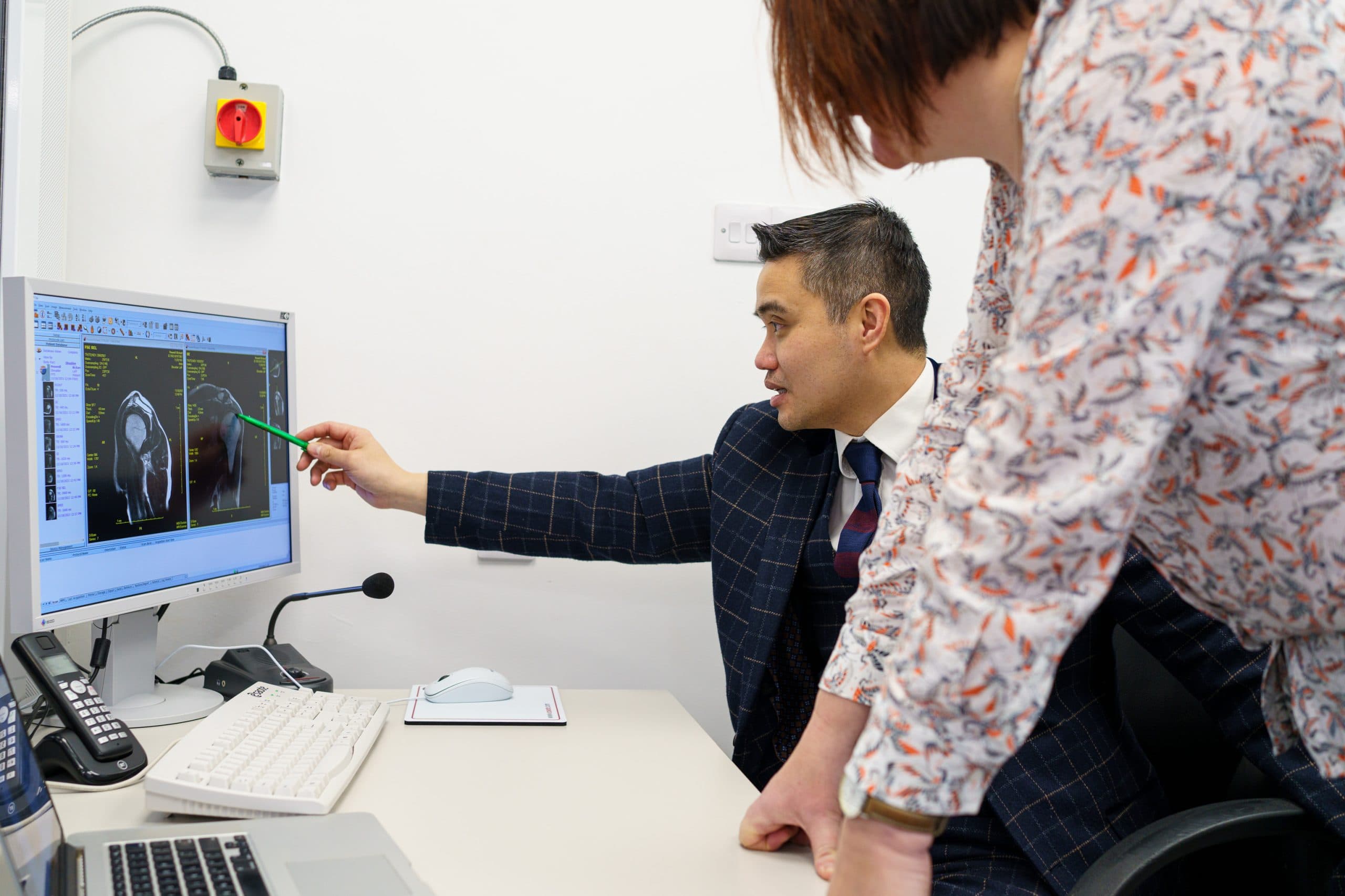
Chondromalacia patellae is the softening and progressive breakdown of cartilage on the underside of the kneecap. It produces anterior knee pain, crepitus (grinding), and difficulty with stairs, squatting, and prolonged sitting. At London Cartilage Clinic, we offer a structured pathway from conservative rehabilitation through to surgical intervention for patients whose symptoms have not responded to non-operative treatment.
 Reviewed byProf Paul Lee MBBch, FRCS (Tr & Orth), PhDLast reviewed 1 May 2026
Chondromalacia is classified into four grades based on the severity of cartilage damage beneath the kneecap. Treatment is guided by grade, symptoms, and how much the condition limits daily activity.
The majority of chondromalacia cases are managed without surgery. A targeted rehabilitation programme addressing muscle balance, patellar tracking, and load management is the foundation of treatment.
At London Cartilage Clinic, we focus on understanding what is driving the cartilage damage, not just treating the symptoms. Correcting the mechanical cause gives any treatment the best chance of lasting success.
You may have more options than you think
At London Cartilage Clinic we follow a structured clinical framework across four areas of treatment. Before recommending a single procedure, we assess which combination of approaches gives you the best outcome.
Protect what you have. Slow degeneration and manage symptoms.
Fix specific damage. Torn tissue, unstable joints, structural problems.
Rebuild lost tissue. Biological treatments that stimulate new growth.
When other options are exhausted. Joint replacement as a last resort.
Explore the full range of treatments available for your joint. Each hub page shows every option we offer, organised by clinical approach.

Chondromalacia patellae refers to the softening and progressive breakdown of the cartilage on the underside of the kneecap. It is graded from I (mild softening) to IV (full-thickness cartilage loss with exposed bone). The condition is closely related to patellofemoral syndrome and is a common cause of anterior knee pain.
Common causes include kneecap malalignment, overuse from repetitive bending activities, direct trauma, and muscle imbalance around the knee. It is frequently seen in runners, cyclists, and patients who do a lot of squatting or stair climbing. It can also develop as part of generalised cartilage degeneration.
Many patients respond well to physiotherapy focused on quadriceps and hip strengthening, activity modification, taping, and anti-inflammatory treatment. Lower-grade chondromalacia often improves significantly with a structured rehabilitation programme. Surgery is considered when conservative measures have not provided adequate relief.
Surgical options include arthroscopic debridement to smooth the damaged surface, microfracture to stimulate biological repair, and in more advanced cases, cartilage regeneration techniques or biological augmentation. The choice depends on the grade of damage, patient age, and activity goals.
Not exactly. Chondromalacia refers specifically to softening of the patellar cartilage, whereas arthritis involves broader joint degeneration. However, advanced chondromalacia (grade III or IV) can progress to patellofemoral arthritis if left untreated, which is why early management is important.
Still have more specific concerns?
Free Discovery CallLondon Cartilage Clinic
Clinical updates, cartilage treatment guidance, and recovery-focused articles from our specialist team.
A practical overview of cartilage repair, injection therapy, rehabilitation planning, and the clinical evidence that guides joint preservation care.
A practical overview of cartilage repair, injection therapy, rehabilitation planning, and the clinical evidence that guides joint preservation care.
A practical overview of cartilage repair, injection therapy, rehabilitation planning, and the clinical evidence that guides joint preservation care.